Our Medical Team
Consult directly with experienced, board-certified ophthalmologists
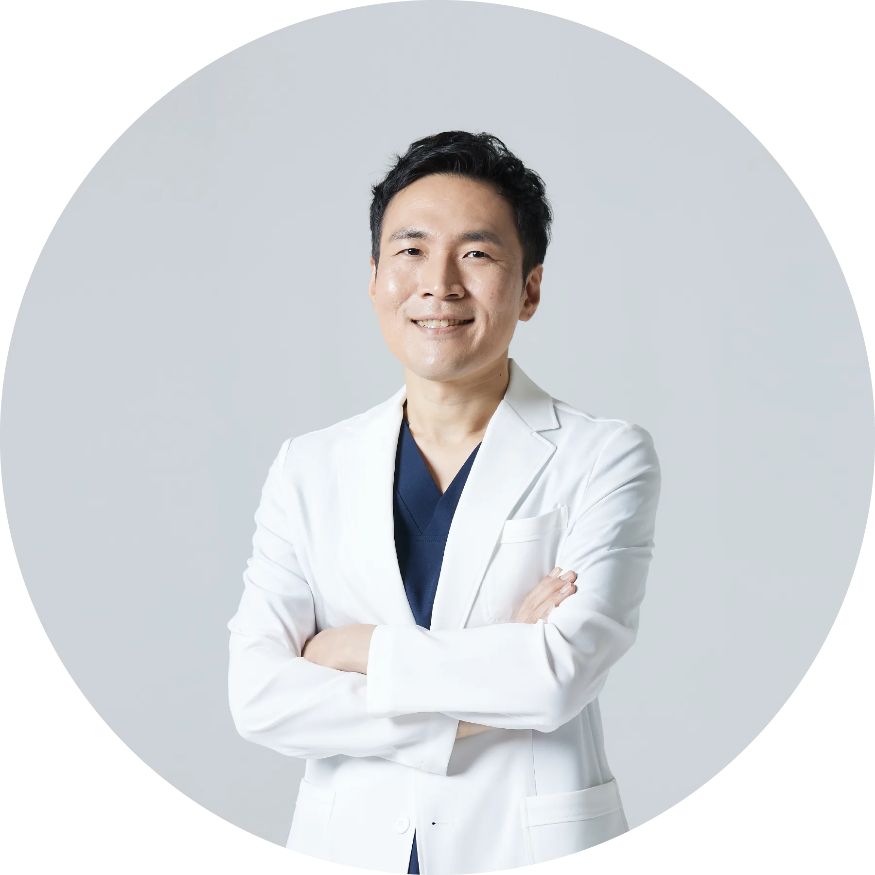
- 서울대학교 의과대학 졸업
- 서울아산병원 인턴 / 안과 전문의
- 서울대학교 안과 전임의(각막, 백내장)
- 前 한길안과병원 진료과장(라식&건성안 센터장)
- 前 카톨릭관동대학교 의과대학 교수, 교육부장

- 서울대학교 의과대학 졸업
- 서울아산병원 안과 전공의 / 각막 임상강사
- 前 충북대학교병원 안과 교수(각막)
- Flaum Eye Institute, University of Rochester 단기연수
- Sickkids Hospital, University of Toronto 연수

- 순천향대학교 의과대학 졸업
- 순천향대학교 대학원 각막 및 외안부 전공
- 서울대학교 안과 전임의(각막, 백내장)
- 前 순천향대학교병원 안과 과장 / 순천향대학교병원 교수
- 대한안과학회(KOS)・미국백내장굴절수술학회(ASCRS)・유럽백내장굴절수술학회(ESCRS) 정회원
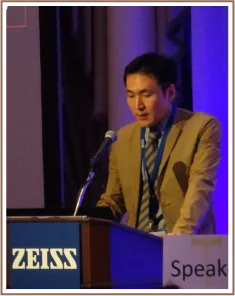
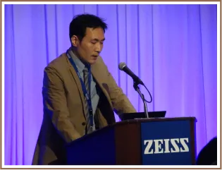
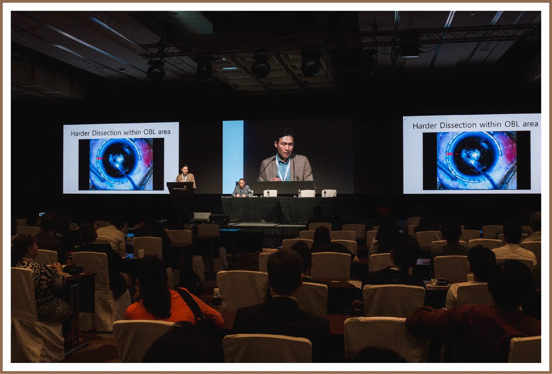
Presented at ESCRS, winning the Best Poster Award; also presented at the ZEISS Asia-Pacific SMILE Symposium. Research on the opaque bubble layer (OBL) that can interfere with SMILE surgery, identifying its risk factors.
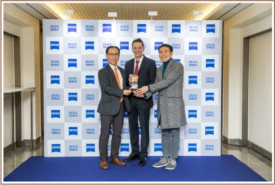
Presented findings on visual quality (clarity) after SMILE surgery — going beyond simple visual acuity.
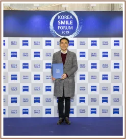
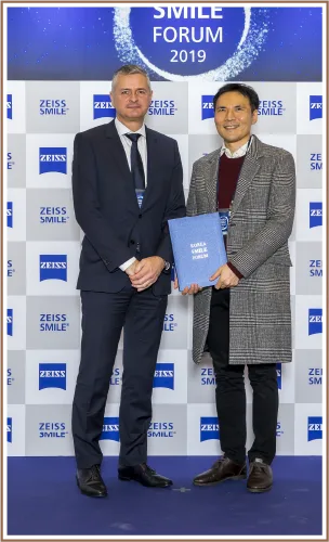
Researched and proposed methods to reduce OBL occurrence; presented at the 2018 Korean Ophthalmological Society conference and the 2019 Tokyo ZEISS Refractive Symposium.
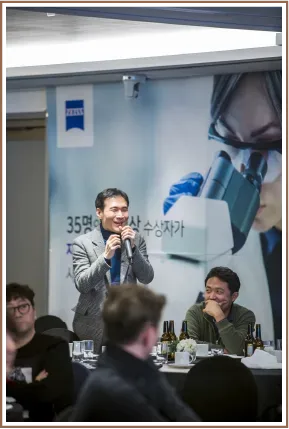

An early adopter of IPL for dry-eye treatment; presented at the international dry-eye society together with the Severance Hospital research team — the first such presentation from Korea.
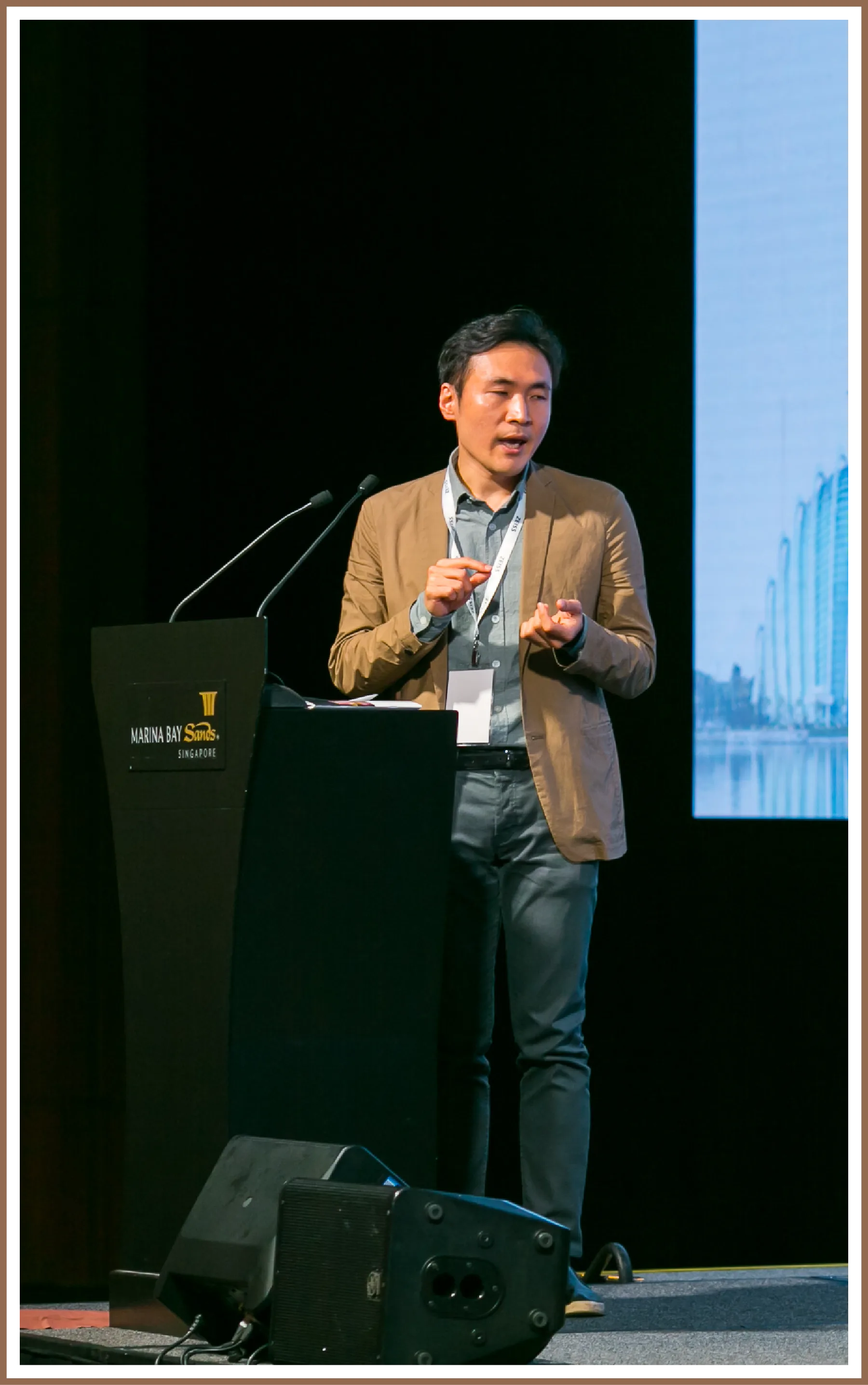
An early adopter of the multifocal IOLs now in widespread use; presented the clinical results at a Korean academic conference.
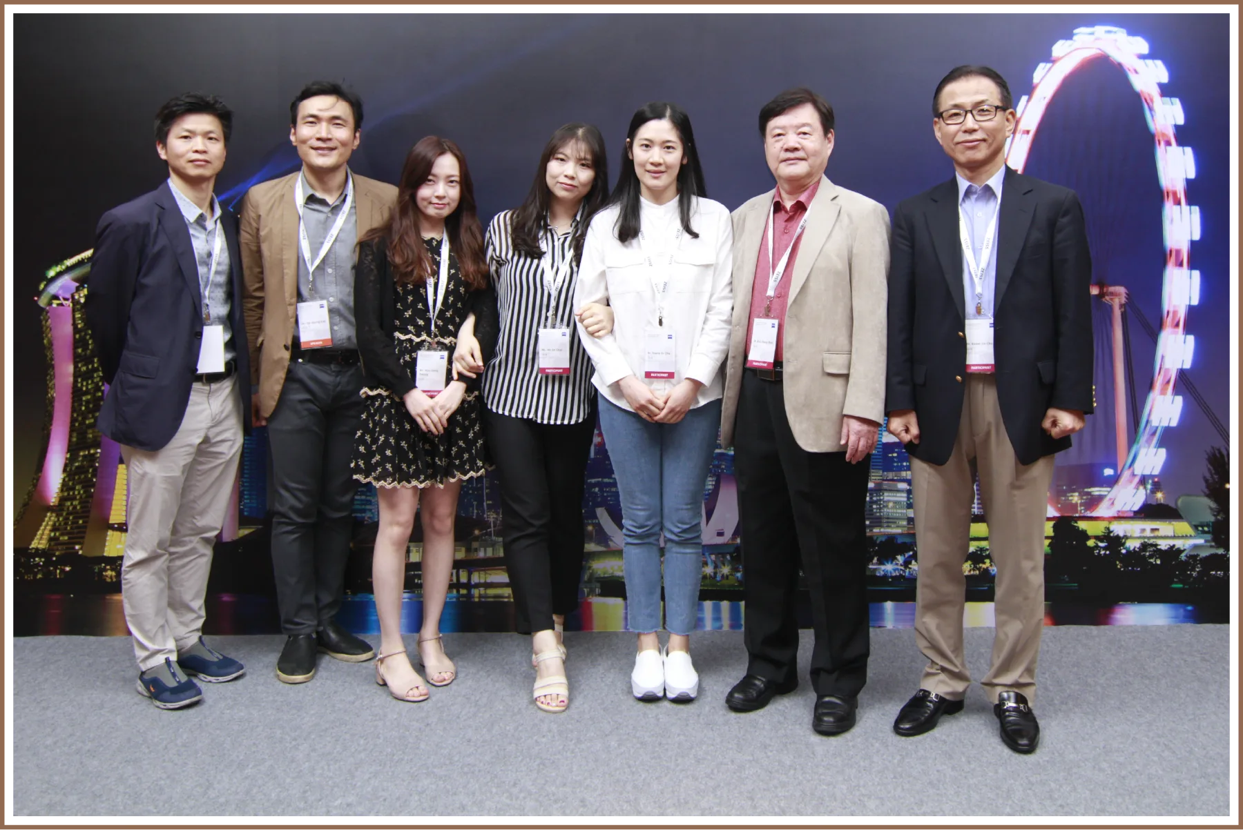
Research on the long-term effects of IPL in dry-eye treatment and how to maintain those effects — the first presentation of its kind on long-term efficacy.
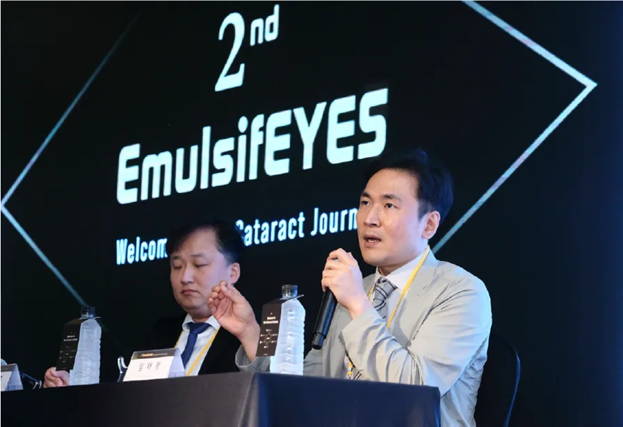
Presented clinical experience with the SS-OCT (Swept-Source OCT) technology in the ARGOS biometer, a device that supports surgical planning and IOL power calculation.
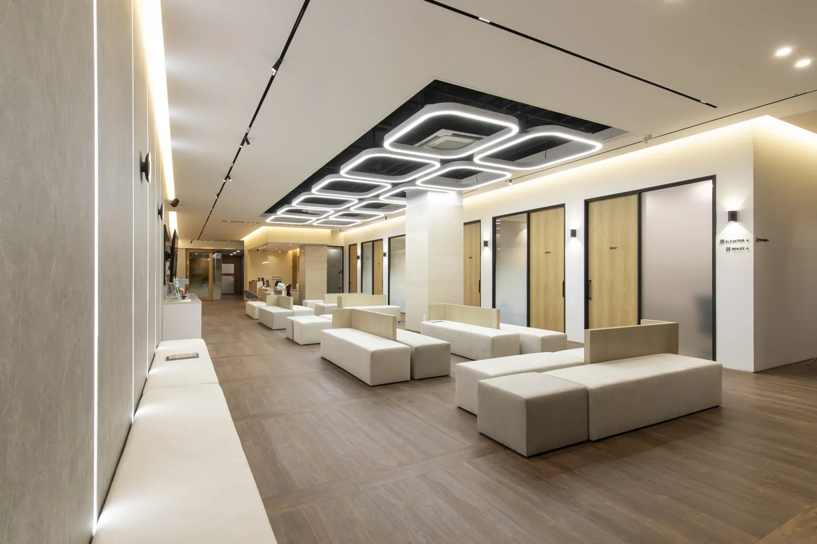
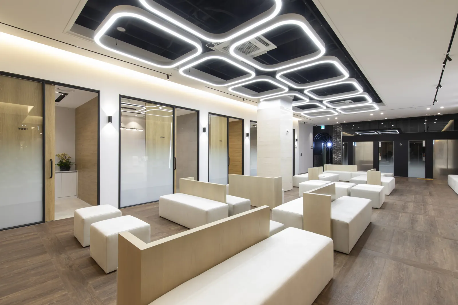
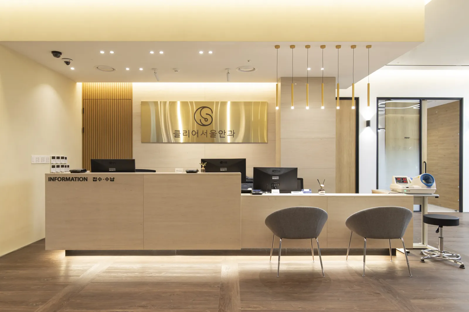
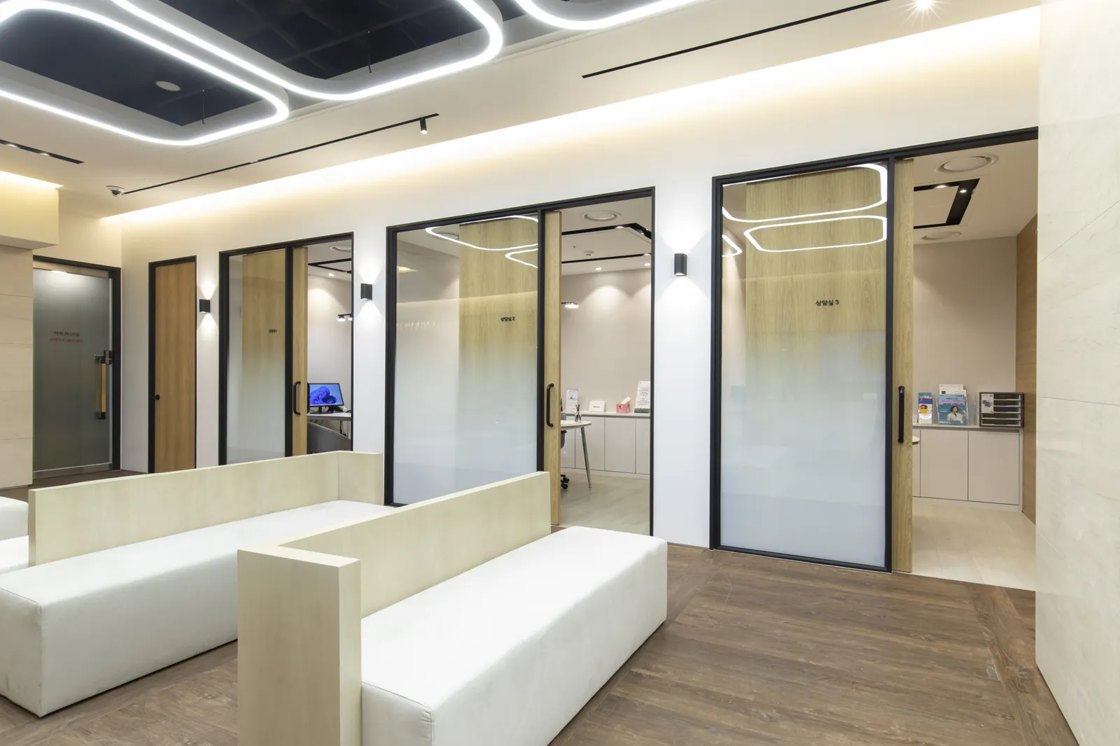
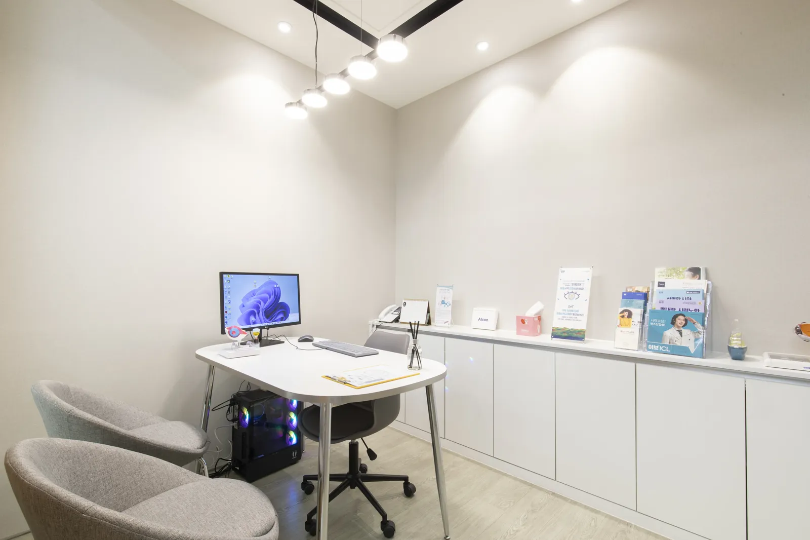
Gangnam, Seoul Eye Clinic — About ClearSeoul
2 min from Gangnam Station Exit 10 — specialists in vision correction, presbyopia & cataract
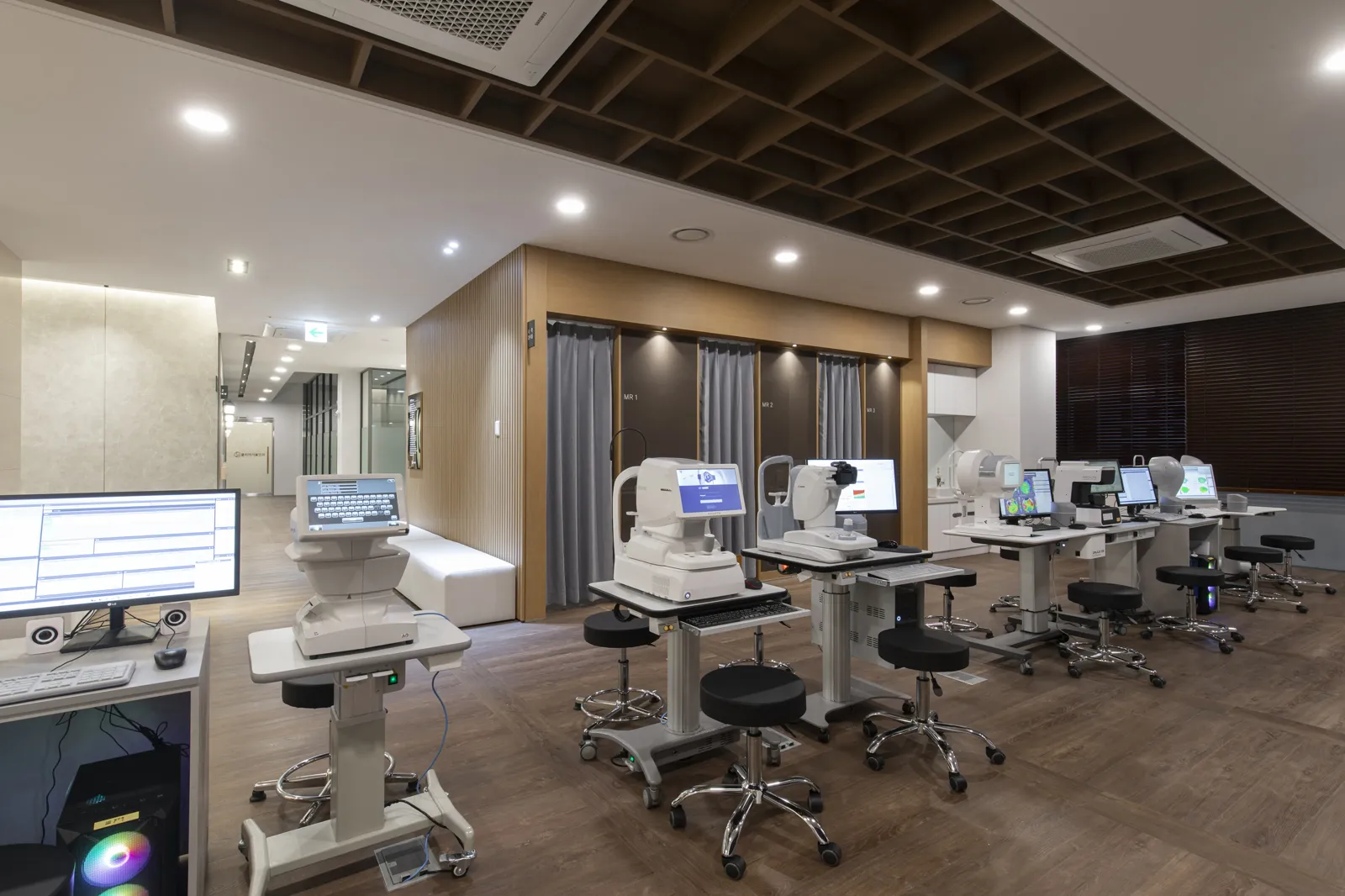
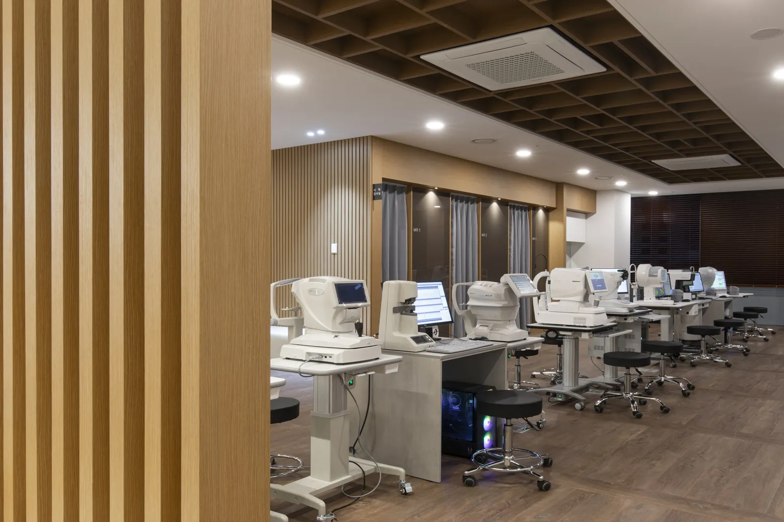
Advanced Medical Equipment
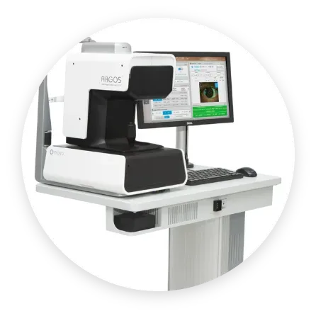
An optical biometer that precisely measures axial length and corneal curvature before cataract and presbyopia surgery to calculate intraocular lens power, supporting accurate lens selection.
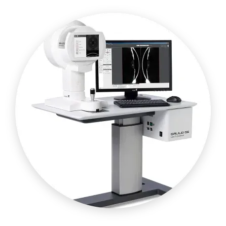
A corneal tomography and topography analyzer that maps the front and back surfaces and thickness of the cornea in 3D, helping assess suitability for vision correction surgery.
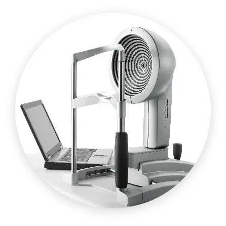
A corneal topographer that precisely measures the curvature of the corneal surface to detect astigmatism and corneal irregularities, providing baseline data for vision-correction planning.
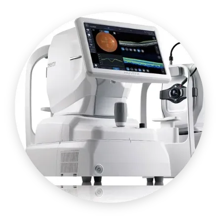
An optical coherence tomography device that captures cross-sections of the retina and optic nerve without contact, supporting early detection of glaucoma and retinal disease.
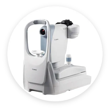
A Canon diagnostic imaging unit that photographs the retina and optic nerve at the back of the eye, used for comprehensive eye-health assessment.
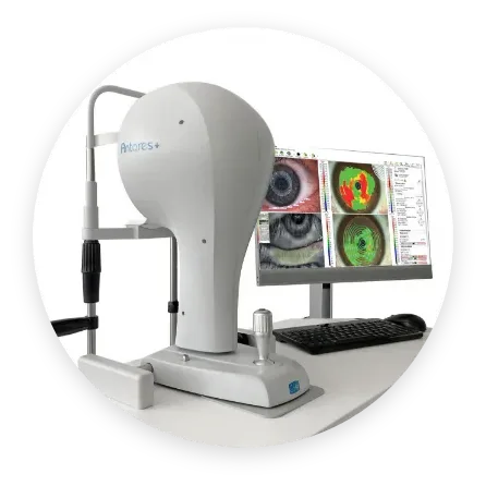
A precision analyzer that visualizes corneal shape and surface condition as color maps, assessing both suitability for vision correction and corneal health.
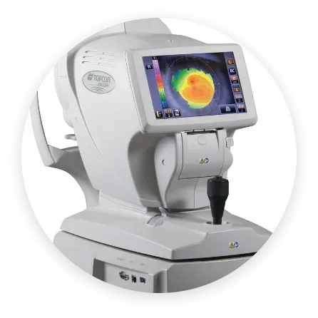
A TOPCON precision diagnostic device that analyzes corneal shape and fundus condition with color maps, used for vision correction and eye-disease screening.
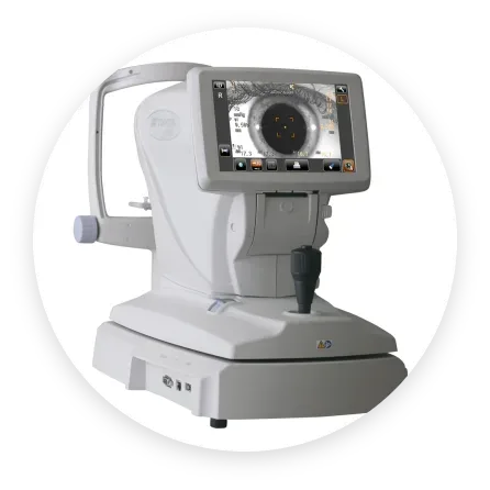
An automated device that quickly measures refractive errors — myopia, hyperopia, astigmatism — and corneal curvature, forming the basis for accurate prescription assessment.
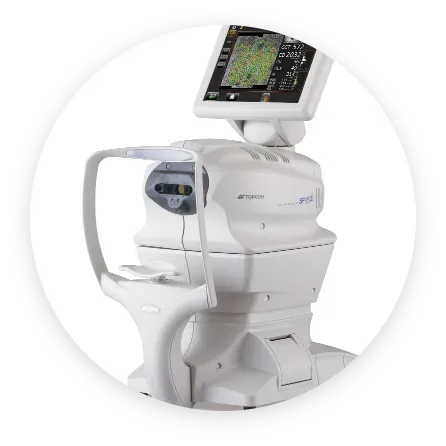
A TOPCON OCT system that captures cross-sectional images of the fine structures of the retina and optic nerve for precise monitoring of glaucoma and retinal changes.
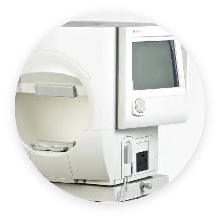
An automated perimeter that measures the range of vision the eye perceives, used to detect visual-field changes from glaucoma and to support regular monitoring.
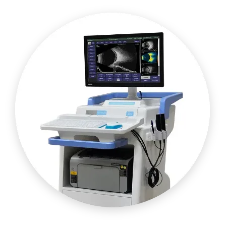
An ophthalmic ultrasound system that measures internal eye structures and axial length, supporting cataract surgical planning and diagnosis of eye conditions.
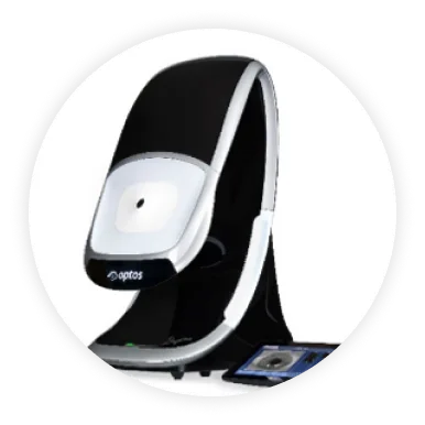
An ultra-widefield imaging device that captures a broad area of the retina in a single, non-contact, non-dilated shot, allowing review of peripheral retinal changes.
Why Choose ClearSeoul Eye Clinic